Lameness Investigations Investigations
We have full facilities for lameness and poor performance investigations.
Lameness and poor performance investigations
Lameness and poor performance investigations can be performed at our practice, where we benefit from a purpose-built trot-up area for walking, trotting and flexion testing.
For more subtle lameness, our ménage allows lunging on a hard or soft surface as well as assessment under saddle or driven.
It is preferable for horses to be ridden by their usual rider, if this is not possible one of our experienced veterinary nurses can ride your horse as part of their lameness investigation.
We do prefer to admit horses to the clinic for lameness investigation rather than carrying these out at yards, especially for more subtle “performance level” cases, as our facilities usually allow for a more accurate and safer diagnosis.
How it works
Normally, the horse will have been assessed at the yard first before an appointment for investigation is made.
If insured, we advise that you notify your insurance company of a likely claim prior to the horse being admitted for investigation, to minimise issues with your claim being processed.
Should you have any queries on insurance cover for lameness investigation, please contact the office on 01306 628222, or the veterinary surgeon in charge of the case.
Once admitted, your horse will be reassessed for stance, muscle symmetry and palpable abnormalities before going on to the dynamic phase of the investigation.
Once the gait has been fully assessed, including riding/driving if necessary, a sequence of nerve blocks will be performed, and the gait reassessed after each one.
These nerve blocks are likely to include blocks into joints or other structures such as tendon sheaths where scrupulous sterility is required, hence the increased safety of carrying these out at the clinic in our treatment rooms.
These nerve blocks allow us, in most cases, to establish the site of lameness and go ahead with imaging of the affected area.
We have diagnostic ultrasound machines and digital radiography that allow us to reach a definitive diagnosis in the majority of cases.
Should further, more advanced investigation be required we will discuss specific options, costs and referral at the time of investigation.
Once a definitive diagnosis has been reached, we can discuss the options for treatment, their costs and the prognosis with you.
Treatment can often be carried out, or at least started, on the same day as the investigation.
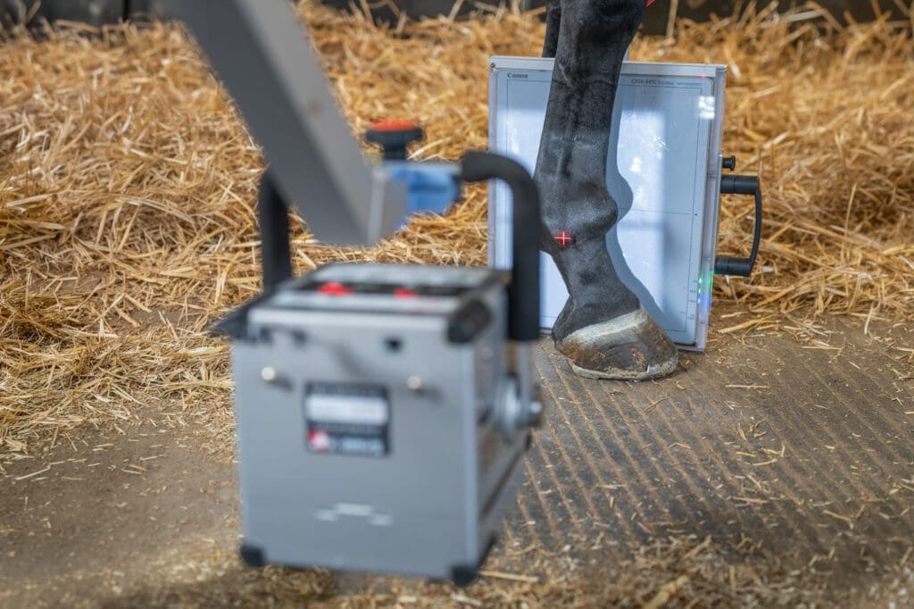
Digital x-ray
Our practice has two digital x-ray machines, one of which operates on a wireless system allowing for x-ray in the field, specifically in an emergency situation where power is not available.
This equipment has revolutionised our approach to possible fractures or laminitis cases which cannot be transported, and increases the service we can provide to clients who do not have access to horse-transport.
Ultrasound scanning
Diagnostic ultrasound allows us to image soft tissues and is most commonly used to assess the reproductive tract in mares, and tendons and ligaments.
However, there are many other uses including examining the abdomen and chest, assessing wounds for foreign bodies, assessing joints, evaluating muscles for damage and examining the eye.
We can image structures up to 25cm beneath the skin.
Shockwave therapy
Extra-corporeal shockwave therapy (commonly referred to as shockwave therapy) Shockwave therapy, is a well established treatment modality for certain orthopaedic issues in the horse.
Structures treated include ligaments, particularly the sacro-iliac, check, collateral and suspensory ligaments, tendons and muscles.
Our shockwave machine is “focused”, which means that it delivers the shockwaves as specifically as possible into the structure to be treated.
The treatment can reduce pain in the affected structure, help to break down scar tissue and can encourage new cells to migrate into the tissue for repair.
The treatment regime usually involves three sessions, each 10 to 14 days apart.
In most cases, the patient will be sedated as the machine is quite loud and creates a strange sensation in the treated area.
Adverse after effects are rarely seen with many of our clients reporting an immediate improvement in their horses’ condition after treatment.
Shockwave can also be used alongside other treatments such as local steroid injections.